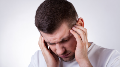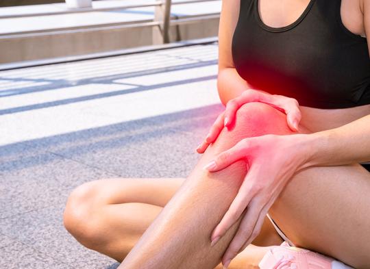
Knee pain is currently one of the most common complaints reported by patients, affecting both young, physically active individuals and older adults struggling with degenerative joint disease.
Causes of knee pain vary widely: from sudden mechanical injuries, through overload related to lifestyle, to chronic inflammatory conditions. Correctly identifying the source of pain is essential in order to initiate effective treatment and avoid permanent functional impairment.
Regardless of whether the pain appeared suddenly after an injury or has been present for years, you can find professional help at our Polish medical clinic. We offer on-site ultrasound scan diagnostics and fast access to experienced orthopaedic specialists in London, who will accurately identify the cause of your symptoms and select an appropriate treatment plan.
Pain location – what does the site of discomfort tell us?
During a medical consultation, an orthopaedic surgeon will always ask their patient to precisely indicate where the pain is most intense. Knee joint is a complex mechanism, and pain rarely spreads across the entire area without a specific reason. The location of discomfort is the first and most important clue, allowing the specialist to narrow down potential diagnose even before imaging tests are performed.
Pain at the front of the knee (around the kneecap)
Pain located at the front of the knee, directly on or just below the patella, most often indicates problems with the knee extensors. If the pain worsens when climbing stairs, squatting, or after prolonged sitting with bent knees (the “cinema-goer’s sign”), it may suggest patellofemoral pain syndrome or softening of the patellar cartilage (chondromalacia). Localised pain slightly below the kneecap is a sign of “jumper’s knee,” i.e. patellar tendonitis. Pain just above the patella is often associated with overload of the quadriceps tendon.
Pain on the sides of the joint (medial and lateral)
Pain on the inner (medial) side of the knee is one of the most common clinical findings. It may suggest damage to the medial meniscus or a strain of the medial collateral ligament (MCL). Early symptoms of degenerative changes also often manifest in this area. Pain on the outer (lateral) side can be misleading. While it may indicate injury to the lateral meniscus, in physically active individuals (especially runners) it’s very often caused by iliotibial band syndrome (ITBS). This pain is sharp, stabbing, and usually appears after running a specific distance.
Pain at the back of the knee (popliteal fossa)
Pain located at the back of the knee is less common but very characteristic. It is often caused by a Baker’s cyst, which is a fluid-filled sac that develops as a result of other pathologies within the knee joint (e.g. meniscal damage). Patients may feel a lump behind the knee and a sensation of pressure during full extension or flexion. Pain in this area may also result from tight hamstring or calf muscles.
“Deep” knee pain and instability
Diffuse pain deep inside the joint, accompanied by a feeling that the knee is “giving way” or instability when walking, is a typical symptom of anterior cruciate ligament (ACL) injury. If, however, the pain presents as morning stiffness that eases with movement and is dull and constant, it usually indicates progressing osteoarthritis.
The most common causes of knee pain
Diagnosing knee pain can be challenging, as the same symptom may result from many different factors. In clinical practice, causes are generally divided into two main categories: acute mechanical injuries related to accidents or physical activity, and chronic changes that develop over years due to disease or natural tissue tear and wear.
Mechanical injuries most often affect physically active individuals, but damage to internal joint structures can also occur during something as simple as stepping awkwardly off a kerb.
Ligament injuries
One of the most serious and unfortunately common injuries is rupture of the anterior cruciate ligament (ACL). This usually occurs through a twisting mechanism, when the foot remains planted while the torso rotates suddenly. Patients often hear a characteristic “pop,” followed by immediate swelling and instability of the knee. Strains of the collateral ligaments (MCL and LCL) are also common, though usually less severe, and occur due to blows to the side of the knee.
Meniscal tears
Overuse syndromes
Not all pain results from a sudden accident. In runners, cyclists, and manual workers, common problems include runner’s knee (iliotibial band syndrome) and jumper’s knee (patellar tendonitis). These conditions result from cumulative microtrauma, inadequate recovery, and muscle imbalance, when some muscle groups are overly tight and others too weak, disrupting normal patellar tracking.
Chronic conditions and inflammatory diseases
Pain that gradually increases over months or even years usually has a degenerative or systemic cause. It most often affects older adults but is more often diagnosed in middle-aged patients as well.
Osteoarthritis (gonarthrosis)
This is the most common cause of chronic knee pain. This condition involves gradual wear of the articular cartilage, which loses its smoothness and elasticity. Over time, exposed bone surfaces begin to rub against each other, causing inflammation, pain, and reduced mobility.
Symptoms often worsen with changes in weather and in the morning; patients report “start-up pain” that improves after a few steps and warming up the joint.
Inflammatory diseases (rheumatoid arthritis and gout)
Knee pain may also be a sign of systemic disease. In rheumatoid arthritis (RA), the immune system mistakenly attacks the body’s own tissues, leading to painful swelling and joint destruction. Gout, on the other hand, is associated with the deposition of uric acid crystals in the joint. Gout attacks are extremely painful, appear suddenly (often at night), and are accompanied by redness, warmth, and tension of the skin around the knee.
Baker’s cyst
A common cause of discomfort in the popliteal area is a Baker’s cyst. This is a collection of synovial fluid that forms at the back of the knee as a defensive response to another pathological process within the joint (e.g. meniscal injury or inflammation). The cyst may compress nearby vessels and nerves, causing pain radiating into the calf.
Diagnosis of knee pain
Effective knee pain treatment is impossible without accurately identifying its cause. Due to the joint’s complex structure, pain location alone may be misleading. Symptoms felt in one area may originate from a completely different structure (e.g. hip pain radiating to the knee). The diagnostic process usually consists of three stages: medical history, physical examination, and imaging studies.
Medical history and physical examination
Diagnosis begins with a visit to an orthopaedic surgeon or physiotherapist. Specialist assesses limb alignment (varus or valgus), joint contour (presence of swelling or bruising), and range of motion. A key element is clinical testing, during which the clinician performs specific movements to assess the integrity of individual structures:
- Meniscal tests (e.g. McMurray test): provoke pain or a “click” in cases of meniscal damage.
- Ligament tests(e.g. drawer test, Lachman test): assess joint stability and detect cruciate ligament ruptures (ACL, PCL).
Imaging studies
Modern medicine offers a range of tools to “look inside” the knee. The choice of imaging depends on the suspected diagnosis.
- X-ray: the first-line test, especially after injury (to exclude fractures) and in older patients. X-rays clearly show bony structures and allow assessment of joint space narrowing, the main indicator of osteoarthritis severity. In suspected degeneration, weight-bearing (standing) X-rays are recommended.
- Ultrasound scan (US): an inexpensive, fast, and safe test. It is ideal for evaluating superficial soft tissues such as collateral ligaments, the patellar tendon, and the popliteal area (Baker’s cyst). Importantly, ultrasound is a dynamic examination; doctor can observe tissue during knee movement.
- Magnetic resonance imaging (MRI): currently the most accurate diagnostic method and considered the “gold standard” for assessing intra-articular structures. MRI precisely visualises menisci, cruciate ligaments, articular cartilage, and bone marrow. It is essential before planned surgery (e.g. ligament reconstruction) but, due to cost and waiting times, is rarely used as a first-choice test for minor complaints.
Laboratory tests
If the doctor suspects that knee pain is caused by a systemic disease rather than mechanical injury (e.g. gout or rheumatoid arthritis), blood tests may be required. Key parameters include:
- ESR and CRP – markers of inflammation
- Uric acid– elevated levels suggest gout
- Rheumatoid factor (RF) – helpful in diagnosing rheumatic diseases
In cases of severe swelling of unclear origin, doctor may also perform joint aspiration, drawing synovial fluid with a syringe. Analysis of the fluid helps distinguish haemarthrosis (after injury) from inflammatory or purulent effusion (bacterial infection).
Treatment methods for knee pain
Choice of the treatment depends strictly on the diagnosed cause. A fresh sports injury requires a different approach than advanced osteoarthritis. Modern orthopaedics aims to make surgery a last resort, placing the greatest emphasis on conservative treatment and rehabilitation.
Conservative and home treatment (RICE protocol)
In cases of acute injuries, bruises, or strains, management during the first 24–48 hours is crucial. The gold standard is the RICE protocol (or its newer version, PRICE), aimed at reducing swelling and inflammation.
- (P– Protection
- R – Rest: temporary unloading of the limb, avoiding painful movements
- I – Ice: cold packs (through a towel, not directly on the skin) applied for 15–20 minutes several times a day; cold constricts blood vessels, reducing swelling and bruising
- C – Compression: use of an elastic bandage or compression brace to limit swelling
- E – Elevation: keeping the leg above heart level to facilitate venous and lymphatic drainage
Pharmacotherapy
Medications are mainly used to reduce acute inflammation and relieve pain, enabling the patient to begin rehabilitation.
- Non-steroidal anti-inflammatory drugs (NSAIDs) - available orally (e.g. ibuprofen, diclofenac, naproxen) or as topical gels and creams. Topical forms are safer for the stomach and liver, acting directly at the pain site.
- Intra-articular injections:
- Hyaluronic acid (viscosupplementation) - “lubricates” the joint by improving synovial fluid quality, reducing cartilage friction; commonly used in osteoarthritis.
- Steroid injections are powerful anti-inflammatory drugs injected directly into the joint. They act quickly but do not treat the underlying cause, and frequent use may weaken tendons and cartilage.
- Platelet-rich plasma (PRP) is a modern regenerative method. Rich in growth factors plasma is prepared from the patient’s own blood and injected into the damaged area to stimulate natural repair processes (e.g. in partial ligament tears).
Physiotherapy and rehabilitation
This is the most important element of long-term treatment. Without strengthening the muscular support system, even the best performed surgery may not produce lasting results.
The goal of physiotherapy is to restore full range of motion and improve joint stability.
- Therapeutic exercise: strengthening the quadriceps (the main knee stabiliser) and hamstring muscles. Strong muscles absorb much of the load, relieving stress on ligaments and cartilage.
- Manual therapy: hands-on techniques to mobilise the joint, improve patellar tracking, and release tight soft tissues.
- Physical therapy modalities: treatments such as cryotherapy, magnetic field therapy, laser therapy, or ultrasound, which support tissue regeneration and provide pain relief.
Surgical treatment
Surgery is considered when conservative methods fail or when mechanical damage has no chance of healing on its own (e.g. complete ACL rupture in an active person, a meniscus tear causing joint locking).
- Arthroscopy: a minimally invasive procedure involving insertion of a camera and miniature instruments through small incisions. It allows suturing or partial removal of a meniscus, ligament reconstruction, or cartilage smoothing, with rapid recovery.
- Osteotomy: a corrective procedure in which a bone is cut and realigned to unload the damaged part of the joint (used in varus or valgus deformities).
- Arthroplasty (joint replacement): the final option in advanced osteoarthritis, when the joint is severely damaged and pain prevents walking. It involves removing destroyed joint surfaces and replacing them with artificial components (a prosthesis).
When does knee pain require urgent medical attention?
Most knee pain can be assessed during a routine appointment. However, there are “red flag” situations in which delay may lead to irreversible joint damage or permanent disability. In such cases, immediate action is required, either an emergency department (A&E) visit or urgent orthopaedic consultation.
Mechanical joint locking
Sudden locking of the knee in one position is a particularly alarming symptom. Patient cannot fully straighten or bend the leg and feels physical resistance inside the joint. This usually indicates displacement of a large meniscal fragment (“bucket-handle tear”) or the presence of a loose body wedged between the bones. Forcing the knee straight is prohibited, as it damages cartilage surfaces. This condition almost always requires prompt surgical intervention (arthroscopy).
Massive swelling (haemarthrosis) after injury
If the knee swells rapidly after twisting or impact (within minutes to an hour), with tense, shiny skin, this indicates bleeding into the joint. Such a massive haemarthrosis most often signals serious injury such as ACL rupture or an intra-articular fracture. Blood inside the joint is destructive to cartilage, so urgent diagnostics and often joint aspiration are required.
Signs of infection: fever, redness, warmth
Knee pain accompanied by fever, chills, marked redness and warmth of the skin around the joint may indicate septic (purulent) arthritis. Infection may occur via the bloodstream or following injections or procedures. This is a life-threatening condition (risk of sepsis) and can rapidly destroy the joint. Immediate hospitalisation and intravenous antibiotics are required.
Inability to bear weight and visible deformity
If after an injury the patient cannot stand on the leg even with support, and the knee’s shape is clearly altered (e.g. the patella displaced to the side or abnormal limb angulation), there is a high likelihood of fracture or joint dislocation. Knee dislocation (not to be confused with a sprain) is particularly dangerous, as it involves tearing of the joint capsule and ligaments and may damage the popliteal artery. This can cause pallor, coldness of the foot, and sensory disturbances, requiring immediate vascular intervention to save the limb.
Knee pain – summary
Knee pain is rarely something that can simply be “waited out.” Whether it results from a sudden skiing injury or slowly progressing degenerative changes, it is a clear warning signal from the body. Ignoring symptoms and masking them with painkillers usually leads to further damage to intra-articular structures and prolongs eventual treatment. Early diagnosis is the key to long-term mobility. Often, a short course of physiotherapy or a targeted injection is enough to avoid complex surgery in the future.
At our Polish medical clinic in London, we successfully combine modern treatments (including hyaluronic acid and PRP injections) with professional rehabilitation. Trust our experience and book a consultation today to return to full fitness as quickly as possible.